A Practical Introduction to Working with Medical Image Data
Figure 1
For example, datasets in ITK-SNAP are illustrated in the following
figures.
Figure 2
Figure. Description of ITK-SNAP tools in the
main window using MRI-crop
example datasets for plane and 3D views.
Figure 3
Figure. ITK-SNAP opening image file
Figure 4
Figure. ITK-SNAP main window
Figure 5
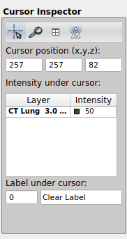
Figure. ITK-SNAP cursor inspector
Figure 6
Figure. ITK-SNAP additional image
Figure 7
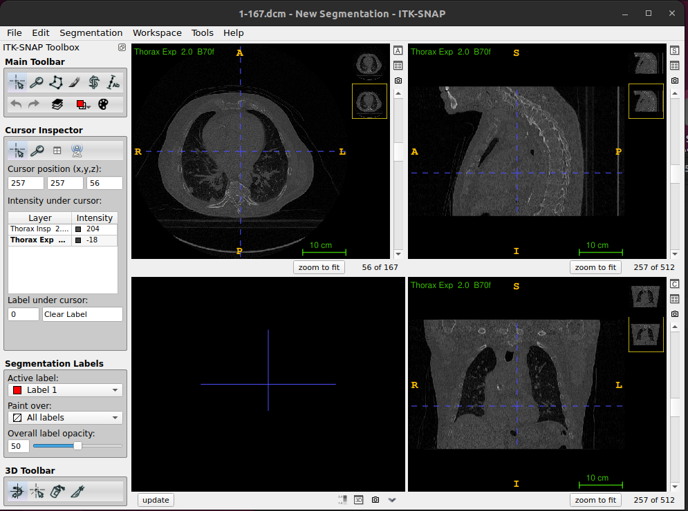
Figure. ITK-SNAP thumbnails, handlding two DICOM
images (
inhale_BH_CT and exhale_BH_CT for
CT-PET-VI-02)Figure 8
Figure. ITK-SNAP thumbnails
Figure 9
Figure. ITK-SNAP semi-transparent overlays
Figure 10
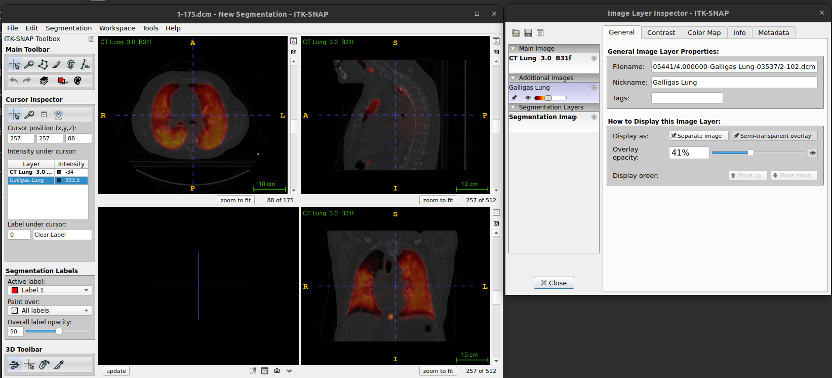
Figure. ITK-SNAP: Layer inspector with general
tab
Figure 11
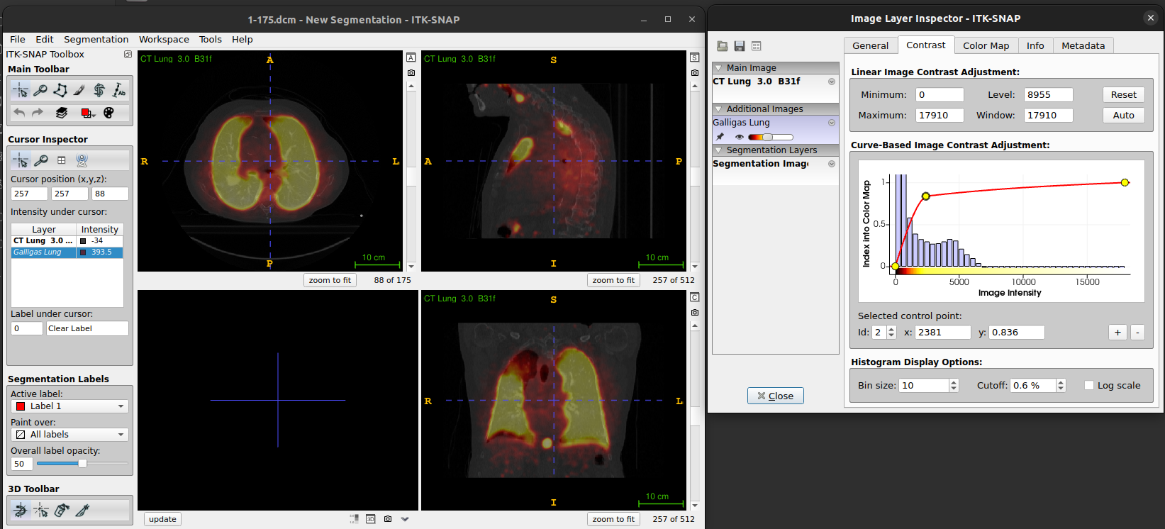
Figure. ITK-SNAP: Layer inspector with contrast
tab
Figure 12
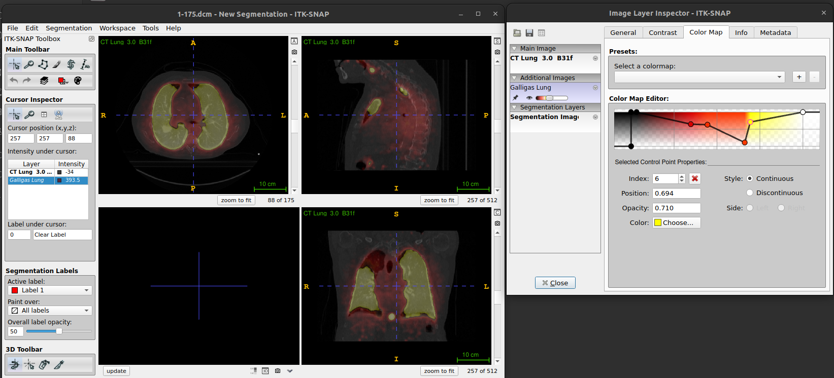
Figure. ITK-SNAP: Layer inspector with color map
tab
Figure 13
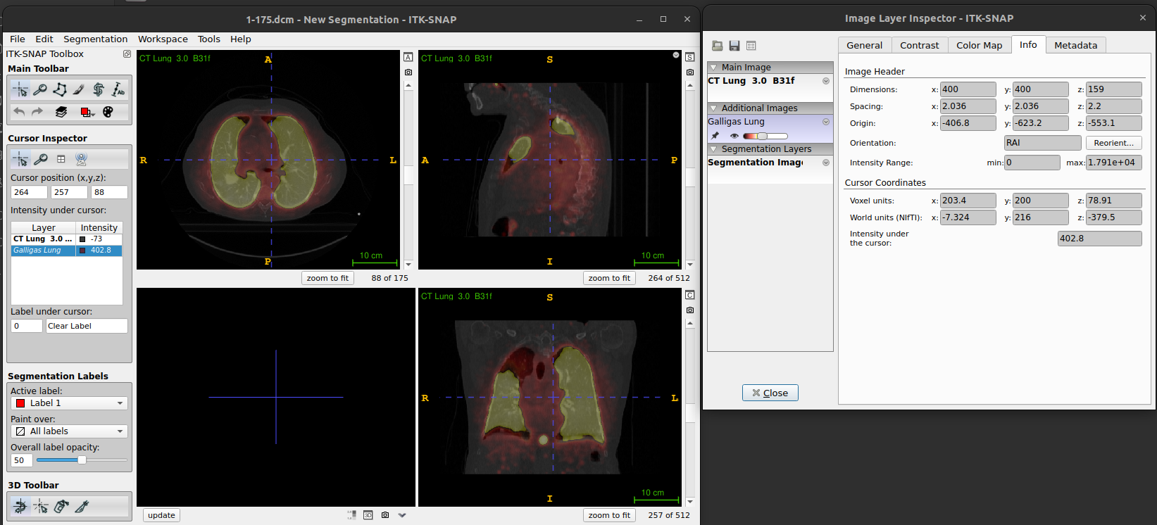
Figure. ITK-SNAP: Layer inspector with info
tab
Figure 14
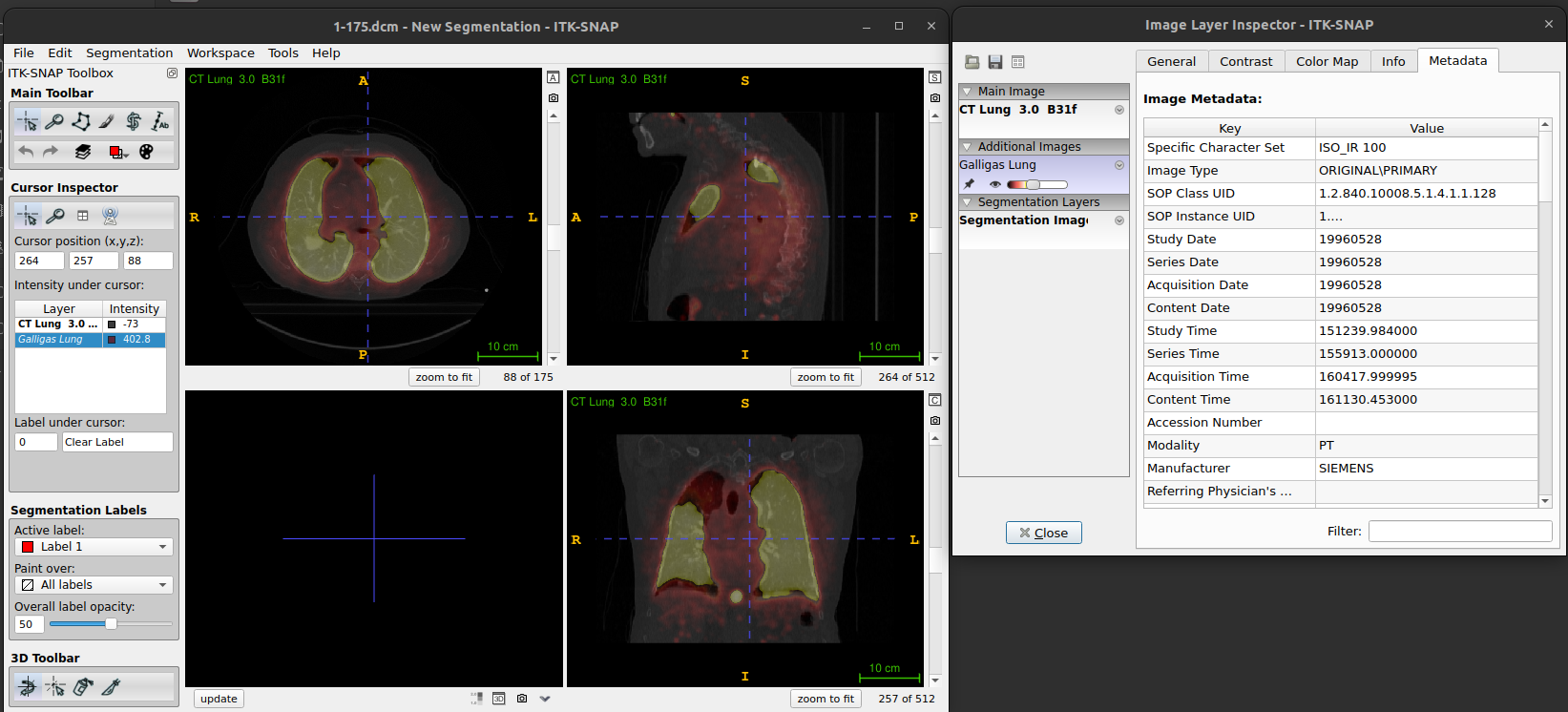
Figure. ITK-SNAP: Layer inspector with metadata
tab
Figure 15
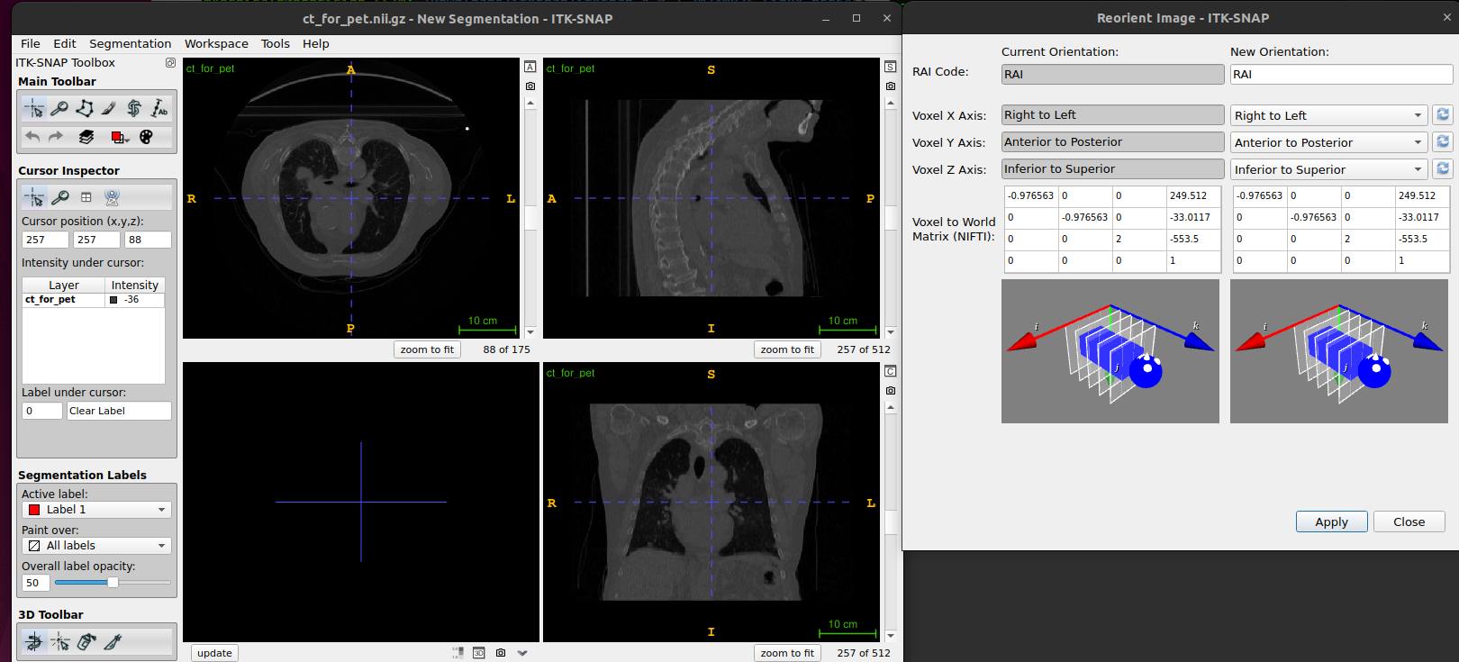
Figure. ITK-SNAP Reorient image
Figure 16
Figure. ITK-SNAP: Metadata for 16-bit integers
vs 32-bit floats
Figure 17
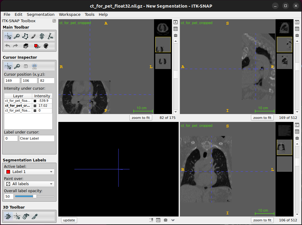
Figure. ITK-SNAP: Cropped image in
ITK-SNAP
Figure 18
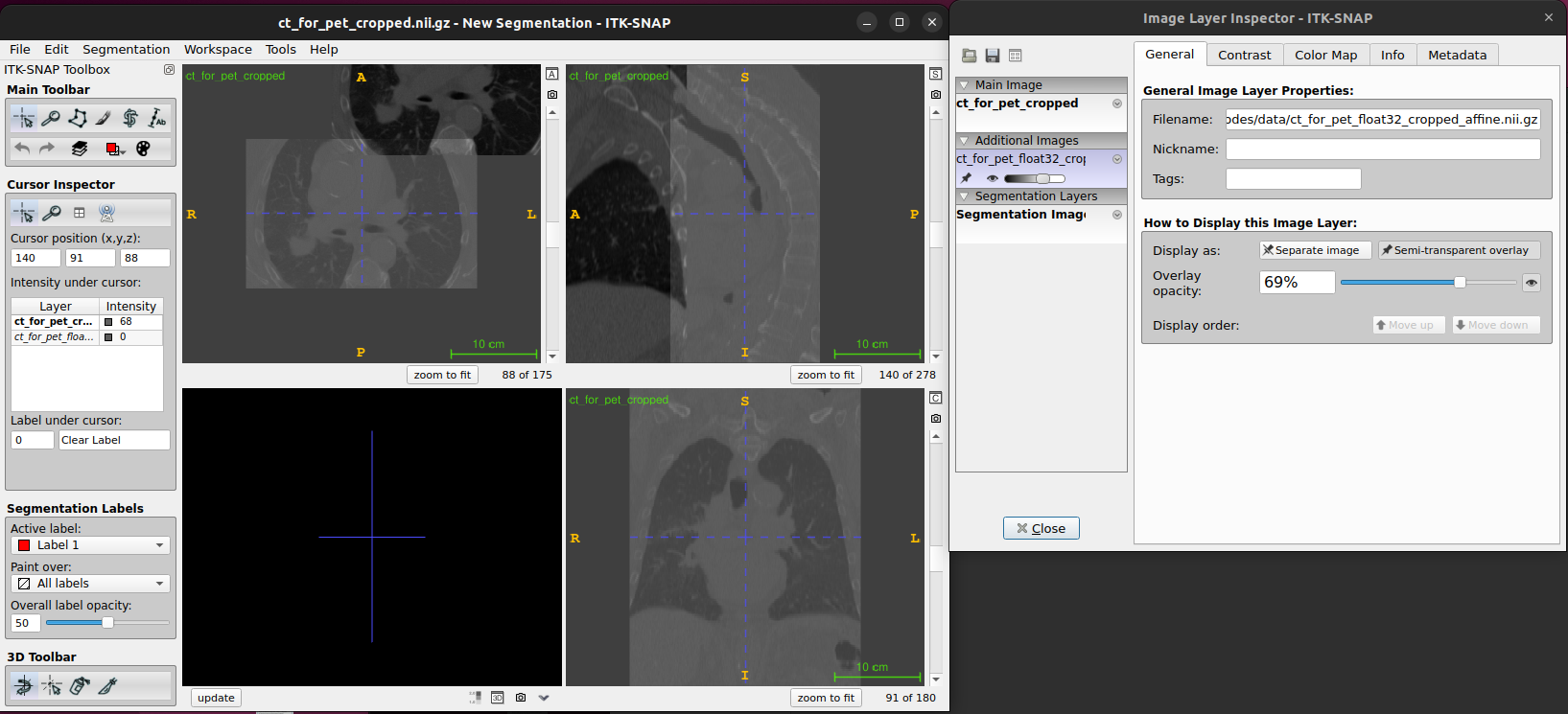
Figure. ITK-SNAP: Cropped image in
ITK-SNAP
Figure 19
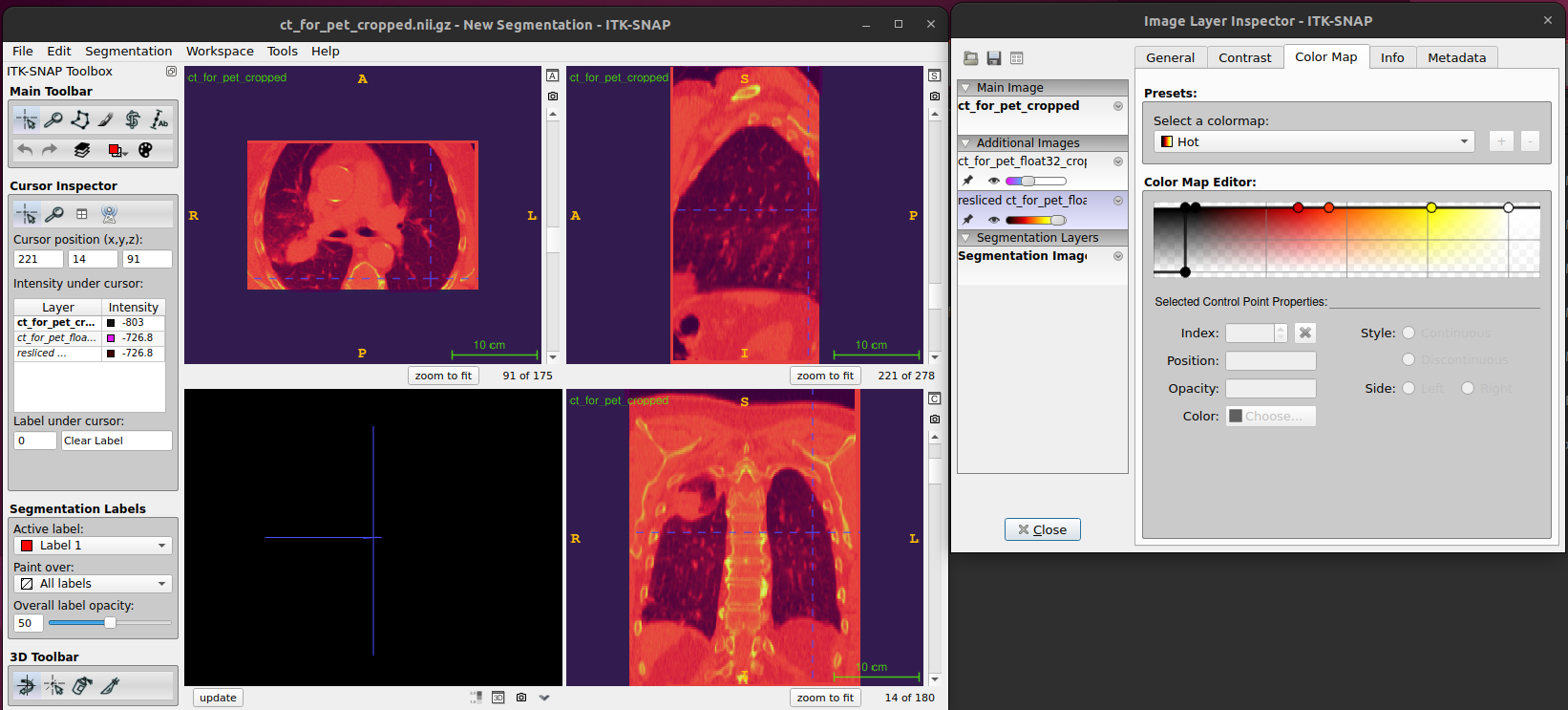
Figure. ITK-SNAP main window
Figure 20
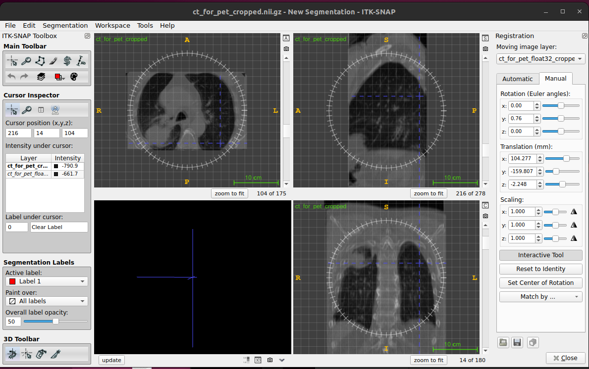
Figure. ITK-SNAP main window
Demons Image RegistrationDemons Image Registration Algorithm with Multi-Pane Display
Figure 1
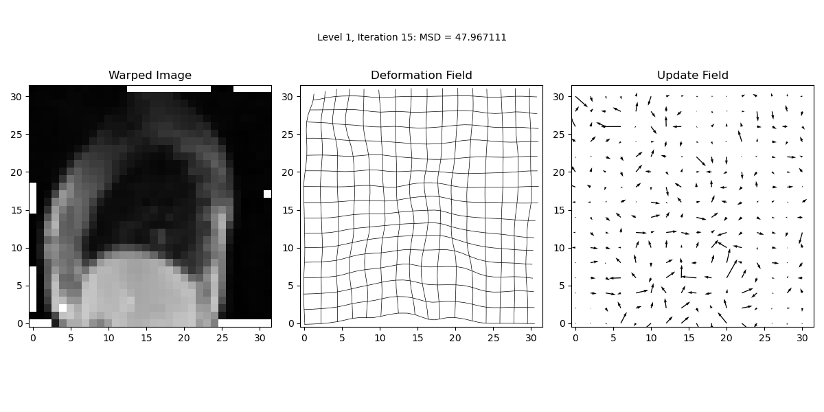
Level 1 Results
Figure 2
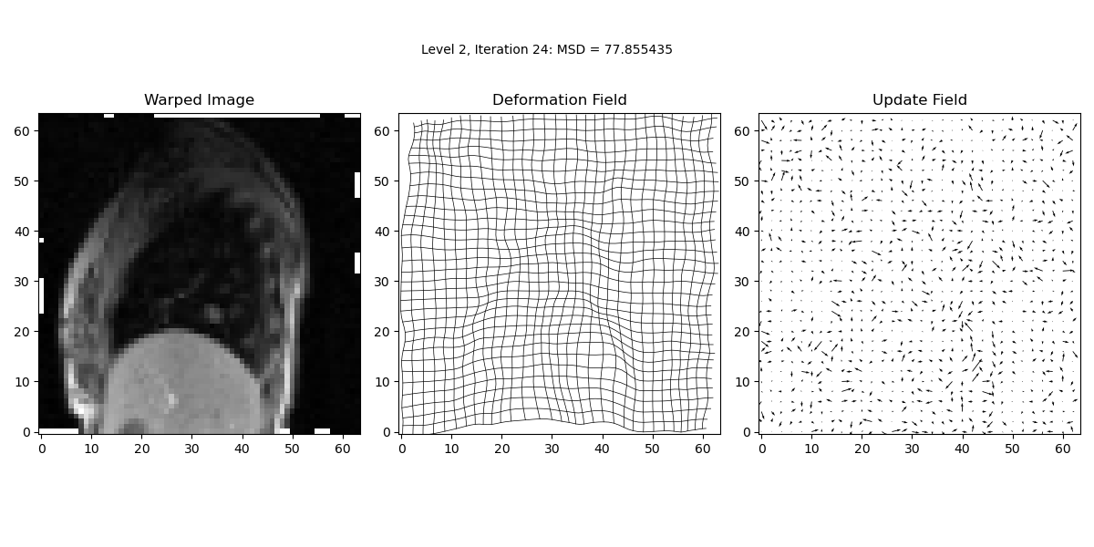
Level 2 Results
Figure 3
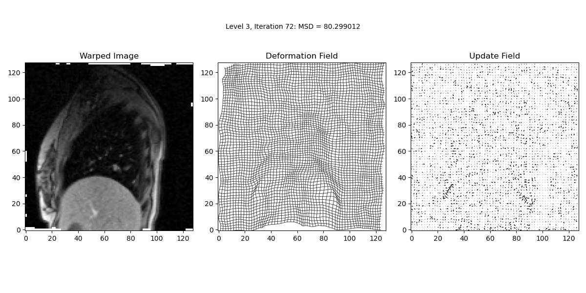
Level 3 Results
